Lupine Publishers Group
Lupine Publishers
Menu
ISSN: 2638-6003
Research Article(ISSN: 2638-6003) 
Incidence of Patella Alta in Anterior Knee Pain- Assessment with Patellar Height Ratio's Volume 1 - Issue 1
Balgovind S Raja1*, Rajib Naskar1, Raunak bhole2 and Anusree Akshay3
- 1Speciality medical officer LT municipal general hospital Sion Mumbai Maharashtra India
- 2Resident KB Bhaba municipal hospital Bandra Mumbai Maharashtra India
- 3MBBS,Mumbai Maharashtra
Received: March 10, 2018; Published: March 16, 2018
Corresponding author: Balgovind S Raja, K B Bhaba Hospital, Maharashtra University of Health Sciences, India
DOI: 10.32474/OSMOAJ.2018.01.000105
Abstract
Background: Altered patellar alignment is associated with anterior knee pain and multitude of conditions that affect the patellofemoral joint. The study aim was to report the incidence of patella altaand patella baja in indian population with anterior knee pain and to investigate whether the normal limits of the patellar height ratios are applicable in indian population.
Methods: Lateral radiographs of 528 knees with anterior knee pain were performed. Patellar heights were measured to calculate the Insall-Salvati (IS), Insall-Salvati Modified (MIS), Blackburne-Peel (BP), Caton Deschamps (CD) ratios.
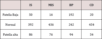
Results: The overall mean LT/LP ratio was 1.03 (SD 0.37) with incidence of 16.2% (86/528) for patella alta with Insall-Salvati. Mean MIS was found to be 1.68 (SD 0.018) and incidence of patella alta as per MIS ratio was 14.3% (76/528). Blackburne-Peel and Caton-Deschamps ratios revealed mean ratios of 0.88 (SD 0.15) and .99 (SD 0.16) and the incidence of patella alta 17.8% with BP ratio and 10.2% with CD ratio respectively.
Conclusion: Incidence of patella alta was found to be 17.8% with BP ratio, 16.2% with IS ratio, 14.3% with MIS ratio and 10.2% with CD ratio. The BP and IS ratio were the most sensitive with MIS ratio being the more specific one and CD ratio the least sensitive.
Keywords: Insall-salvati ratio; Modified insall-salvati; Blackburne-peel; Caton deschamps; Patellar alignment
Introduction
Anterior knee pain remains one of the commonest musculoskeletal complaint with patients, and evaluation of patellar height is done commonly as many conditions are associated with abnormal patellofemoral relationship. The lever arm function of the patella plays an important role in knee extensor mechanism and improves the quadriceps strength by 30-50% [1]. The joint reaction force of the patellofemoral joint varies with the patellar height. A high riding patella or patella alta is associated with patellofemoral malalignment and a reduced patellofemoral contact often leading to patellofemoral pain or instability [2-3]. A low riding patella or patella baja is often associated with limited knee range of motion, patellofemoral arthritis and Osgood Schlatter disease [4]. Numerous methods of patellar height have been described in the literature: Blumensaat [5] Insal-Salvati [6], modified Insal-Salvati [7], Blackburne-peel [8], Caton-Deschamps [9], DeCarvalho [7] and Koshino [10]. These ratios are based on the ratio of the patellar length to the reference point from the tibia. A very few studies have compared the different indices for the patellar height analysis regarding their reproducibility and reliability.
The study objective was to analyse the commonest methods for measuring the patellar height in patients with anterior knee pain and the aim of this study was to report the incidence of patella alta and patella baja and investigate whether the patellar height ratios have significant variations in adult indian population in which sitting on the ground, kneeling, and squatting is common.
Materials and methods
528 lateral x-rays of the knee (212 male and178 females) were collected between the time period August 2017 to February 2018 from patients with anterior knee pain, with difficulty or pain on squatting or sitting. Patients with underlying knee pathologies, knee deformities and knee surgery were excluded. Institutional review board approval was obtained prior to conducting the study. Lateral radiographs of the knee set in 30° of flexion were taken with assistance of a goniometer. 30° of flexion of knee results in better visualization of the tibial insertion. Radiographs were perpendicular to the film and centered on the joint at a distance of 100cm.
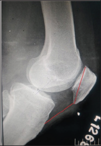
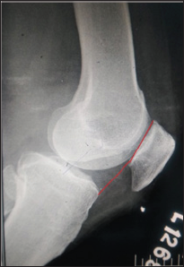
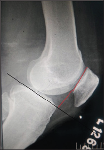
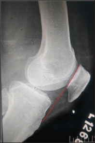
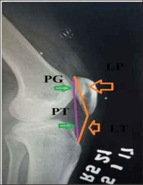
The measurements were made with the digital imaging software (Radiant diacom viewer) by a single experienced physician. The patella length was measured from the superior articular pole to the inferior non articular pole (Figure 1). The distance from the origin of the patellar tendon at the inferior pole of patella to its insertion on the tibial tubercle was taken as the patellar tendon length. The Insall-Salvati index is calculated as ratio of LT/LP, where LT is the length of the patellar tendon and LP is the patella length (Figure 1). The Blackburne-Peel index (Figure 2) is calculated as PP/PG where PP is the perpendicular height of the distal part of the joint surface of the patella to a line projected anteriorly to the surface of the tibial plateau and PG is the length of the articular surface of patella. The ratio PTG/PG, in which PTG is the distance from the lower edge of the articular surface of the patella to the anterosuperior angle of the tibia, and PG is the length of the articular surface of the patella is calculated as Caton-Deschamps index (Figure 3). The Modified Insall-Salvati index consists of the ratio PT/PG, wherein PT is the distance from the lower edge of the joint surface of the patella to the insertion of the posterior or deep surface of the patellar tendon in the tibial tubercle, and PG is the length of the joint surface of the patella (Figure 4).
Results
The overall mean LT/LP ratio was 1.02 (SD 0.37). Comparison between genders revealed that the mean LT/LP ratio was higher in males than females with a mean of 1.04 (SD 0.29) and 1.01 (SD 0.46), respectively (Table 1 & 2). Using criteria of defining abnormal patellar position (1.00±20%) based on Insall's study, the overall incidence was 16.2% (86/528) for patella alta (Tables 3 & 4). The mean PT/PG ratio was 1.68 (SD 0.018). The MIS ratio was higher in males with mean of 1.70(SD 0.29) than females with a mean of 1.66 (SD 0.29). The incidences of patella alta as per MIS ratio were 14.3% (76/528). Blackburne -Peel and Caton- Deschamps ratios revealed mean ratios of 0.88 (SD 0.15) and .99 (SD 0.16). Males were found to have a higher mean in both the ratios compared to the females (BP ratio: 0.89, SD 0.12 and CD ratio: 1.0, SD 0.18 in males and BP ratio: 0.88, SD 0.05 and CD ratio: 0.98, SD 0.06 in females). The incidence of patella alta were 17.8% and 36% with BP ratio and CD ratio respectively.
Discussion
Vast majority of the studies in literature are often quoted with the data obtained from the Caucasian population. There are none, if at all few studies of patellar height being done in Asian population. There are morphological changes in patella of the Caucasian and the Asian population which make the patellar height ratios even more significant. The present study is an observational study done with aim of assessing the patellar alignment in anterior knee pain patients.
The position of the patella has an important role on patellofemoral function. Abnormalities in patella position have thus been associated with anterior knee pain and many extensor mechanism disorders. While many techniques have been developed to measure patellar position such as the Blacburne's ratio, the Insall Salvati ratio still remains one of the most popular, largely because it is easy to use and remember [7,8] .Despite its popularity, recent studies have suggested that the current normal ranges should be extended, as the ratios may not be applied to ethnicities outside western regions [11,12].
The Insall-Salvati (IS) method uses the length of the patellar ligament in relation to the length of the patella6. The patellar morphology and morphological differences in the anterior tuberosity of the tibia (ATT) directly affect the measurements made using this method. Grelsamer and Meadows [7] developed the modified Insall-Salvati (ISM) method based on the length of the joint surface. Difficulty in identifying this parameter is considered to be the main measurement bias. The Modified Insall -Salvati ratio has its advantages over the IS ratio that it can efficiently find out the patella alta in patients with small patellar articular surface which is not taken into account in IS ratio (Figure 5). Digital radiography seems not to present greater details for this anatomical reference. The Blackburne-Peel (BP) method exchanges the reference point of the ATT for the joint surface of the tibial plateau, while keeping the joint surface of the patella. Although Berg et al. [13] found that this was the most accurate and reproducible method in conjunction with the IS index, and Seil et al. [13] ranked it as the second most reproducible method in conjunction with the IS index, we did not obtain similar results in our analysis, such that it was only better than the ISM index. Lack of definition of the reference line of the tibial plateau, such as which condyle to use as the reference, or whether this line runs parallel to the joint surface or perpendicular to the long axis of the tibia, contributes towards lower concordance with this method. The method of Caton et al. [2], which uses the joint surface of the patella and the angle of the tibial plateau as references, also presents difficulty regarding identification of the joint surface, as well as presenting a certain amount of variability in defining the angle of the tibial plateau. Despite these factors, this method was the one that showed greatest concordance in the study by Seil et al. [13].
The mean IS ratio was 1.02(SD 0.37) with the incidence of patella alta being 16.2%. The incidence of patella alta calculated by height ratios was found to be lesser than that of the published literature. Out of the ratios analysed, it ws found out that the IS ratio and BP ratio had the highest sensitivity for patella alta wherein the CD ratio had the lowest sensitivity. The difficulty in analysing the anatomy of the proximal tibia may be attributed to the decreased sensitivity of CD ratio. MIS ratio is helpful in situations where in the patellar articular length is decreased compared to that of the length of patella (Figure 5). The study had the most sensitive ratio as BP ratio followed with IS ratio, MIS ratio and lastly CD ratio which is comparable to the study by Seil et al. [13]. It was interesting to note that, patella baja were present in a proportion of patients with knee pain signifying the requirement of understanding the knee biomechanics and further evaluation of the patellofemoral joint for the articular cartilage. Patella alta was found to have a incidence of 16.2% as per Insall- Salvati ratio and needs to be kept in mind in paients with anterior knee pain.
References
- Krevolin JL, Pandy MG, Pearce JC (2004) Moment arm of the patellar tendon in the human knee. J Biomech 37(5): 785-788.
- Caton HJ, Dejour D (2010) Tibial tubercle osteotomy in patello-femoral instability and in patellar height abnormality. Int Orthop 34(2): 305309.
- Grana WA, Kriegshauser LA (1985) Scientific basis of extensor mechanism disorders. Clin Sports Med 4(2): 247-257.
- Aparicio G, Abril JC, Calvo E, Alvarez L (1997) Radiologic study of patellar height in Osgood-Schlatter disease. J Pediatr Orthop 17: 63-66.
- Blumensaat C (1938) Die Lageabweichungen und Verrenkungen der Kniescheibe. Ergebnisse der Chirurgie und Orthopadie 31: 149-223.
- Insall J, Salvati E (1971) Patella position in the normal knee joint. Radiology 101(1):101-104.
- Grelsamer RP, Meadows S (1992) The modified Insall-Salvati ratio for assessment of patellar height. Clin Orthop Relat Res 282:170-176.
- Blackburne JS, Peel TE (1977) A new method of measuring patellar height. J Bone Joint Surg Br 59(2): 241-242.
- Caton J, Deschamps G, Chambat P, Lerat JL, Dejour H (1982) Patella infera. Apropos of 128 cases. Rev Chir Orthop Reparatrice Appar Mot 68(5): 317-325.
- Koshino T, Sugimoto K (1989) New measurement of patellar height in the knees of children using the epiphyseal line midpoint. J Pediatr Orthop 9(2): 216-218.
- Leung YF, Wai YL, Leung YC (1996) Patella alta in southern China. A new method of measurement. Int Orthop 20(5): 305-310.
- Phillips CL, Silver DA, Schranz PJ, Mandalia V (2010) The measurement of patellar height: a review of the methods of imaging. J Bone Joint Surg Br 92(8): 1045-1053.
- Seil R, Muller B, Georg T, Kohn D, Rupp S (2000) Reliability and interobserver variability in radiological patellar height ratios. Knee Surg Sports Traumatol Arthrosc 8(4): 231-236.

Top Editors
-

Mark E Smith
Bio chemistry
University of Texas Medical Branch, USA -

Lawrence A Presley
Department of Criminal Justice
Liberty University, USA -

Thomas W Miller
Department of Psychiatry
University of Kentucky, USA -

Gjumrakch Aliev
Department of Medicine
Gally International Biomedical Research & Consulting LLC, USA -

Christopher Bryant
Department of Urbanisation and Agricultural
Montreal university, USA -

Robert William Frare
Oral & Maxillofacial Pathology
New York University, USA -

Rudolph Modesto Navari
Gastroenterology and Hepatology
University of Alabama, UK -

Andrew Hague
Department of Medicine
Universities of Bradford, UK -

George Gregory Buttigieg
Maltese College of Obstetrics and Gynaecology, Europe -

Chen-Hsiung Yeh
Oncology
Circulogene Theranostics, England -
.png)
Emilio Bucio-Carrillo
Radiation Chemistry
National University of Mexico, USA -
.jpg)
Casey J Grenier
Analytical Chemistry
Wentworth Institute of Technology, USA -
Hany Atalah
Minimally Invasive Surgery
Mercer University school of Medicine, USA -

Abu-Hussein Muhamad
Pediatric Dentistry
University of Athens , Greece

The annual scholar awards from Lupine Publishers honor a selected number Read More...


