Lupine Publishers Group
Lupine Publishers
Menu
ISSN: 2637-4692
Research Article(ISSN: 2637-4692) 
Quality of Root Canal Filling in Manually and Rotary-Filed Single-Rooted Teeth Undergoing Apicectomy in Tertiary Care Hospital of South Punjab Volume 5 - Issue 3
Muhammad Athar Khan1* and Shamsa Nazar2
- 1Department of Oral & Maxillofacial Surgery, Bakhtawar Amin Medical and Dental College, Pakistan
- 2Department of Community Dentistry , Bakhatwar Amin Medical and Dental College, Pakistan
Received:July 07, 2023; Published:July 18, 2023
Corresponding author:Muhammad Athar Khan, Assistant Professor, Department of Oral & Maxillofacial Surgery Bakhtawar Amin Medical and Dental College, Multan, Pakistan
DOI: 10.32474/MADOHC.2023.05.000225
Abstract
Objective: This study aimed to determine if there was a significant difference in root canal obturation quality between singlerooted
teeth produced using a manual approach and those prepared using a rotational method, as determined by intraoral periapical
radiographs.
Study Design: Randomized control trial.
Place and Duration: This randomized study was conducted at Bakhtawarmin Medical and Dental College during the period
from April 2022 to September 2022.
Methods: A total of 80 patients of both genders were presented. All the included cases had single rooted teeth. Age, gender,
and residence were some of the detailed demographic information that was documented after getting informed written consent.
Patients were equally divided in two groups. Group I received manual technique in 40 cases while group II received rotary system.
Outcomes among both groups were recorded in terms of quality of root canal filling. SPSS 23.0 was used to analyze all data.
Results: There were 45 (56.3%) females and 35 (43.7%) males. The mean age of the patients in group I was 33.3±8.35 years
and in group II mean age was 34.9±7.23 years. Among 40 cases of group I, 22 (55%) patients had urban residency and in group II
19 cases had urban residency. We did not find any significant difference in root canal filling and homogeneity among both groups.
Group II showed a significantly good results of T score as compared to group I with p value <0.005.
Conclusion: According to the results of this research, the rotary approach yielded higher quality obturations in terms of taper
and overall quality than did the traditional method. Despite this, there was no difference between rotary and manual instrumentation
in terms of the radiographic technical quality of the root canal obturation with respect to length and density.
Keywords:Obturation quality; root canal treatment; manual technique; rotary technique; quality; root canal filling; manually; rotary-filed; single-rooted teeth; apicectomy; tertiary care hospital; South Punjab
Mini Review
Root canal filling is a critical procedure that aims to remove infected tissue from the root canal and fill it with a biocompatible material to prevent further infection. Apicectomy is a surgical procedure that involves the removal of the tip of the tooth’s root, which is then sealed with a filling material. The success of an apicectomy largely depends on the quality of the root canal filling. In this article, we will discuss the quality of root canal filling in manually and rotary-filed single-rooted teeth undergoing apicectomy in a tertiary care hospital of South Punjab. The pulp tissue is directly affected by extensive caries in young children, producing pain and discomfort. Necrotic baby teeth are more likely to be saved thanks to pulpectomy, which helps children avoid bad oral hygiene practices and the need to move other teeth to make room [1]. An accurate diagnosis of the pulp state is necessary for determining the best course of therapy for each individual patient. In order to determine the health of the pulp, it is required to do a thorough clinical examination, pain history, high-quality periapical radiograph, and pulp sensitivity testing with low-temperature cold testing [2]. Clinical decision-making should be aided by radiographic diagnosis, which enables for evaluation of caries severity and commitment to supporting tissues. What kind of projection, between bitewing and periapical, is best will depend on the extent of the caries. When it comes to treating the pulp, periapical radiographs are the gold standard since they provide crucial information about the tooth’s overall affectation, bone health, root structure, and even the location of the permanent tooth’s germ. When evaluating pulpitis, it is not appropriate to utilize CBCT on a regular basis. Hemostasis, or the absence thereof, during pulpal therapy will serve as the last evidence of pulpal inflammation [3]. Radiographic images are typically used to assess the quality of endodontic obturation. Furthermore, clinical factors required for obtaining an acceptable root canal obturation can be discovered during the root canal preparation and obturation phases of therapy [4,5]. The presence of voids in the root filling material, the taper of the canal, and the length of the filler material in relation to the radiographic apex are all factors that contribute to the determination of the root filling’s technical quality. To this day, radiographic evaluation [6] has served as the cornerstone of methodologies used to evaluate the RCT’s viability from a technical standpoint.
Root fillings that are placed within 0-2 mm of the radiographic apex had a decreased probability of developing post-treatment disease [7]. This is in comparison to root fillings that are positioned farther than 2 mm away from the apex of the tooth. Root canal treatment (RCT) results have been demonstrated to be substantially impacted by the length of the root filling in relation to the radiographic apex, with studies finding 87-94% healing rates connected with root filling ending 0-2 mm from the radiographic apex. This has been proved to be the case. Lower healing rates were seen for both short root fillings (those that stopped less than 2 mm from the radiographic apex) and long fillings (those that protruded beyond the apex). [8]. One of the features of the prognosis for endodontic treatment is the quality of the shutter. The current gold standard for gauging the quality of endodontic therapy is the periapical radiographic examination. Root canal filling radiography volume, homogeneity, and taper are used to evaluate the success of endodontic treatment. Several research including undergraduates, graduates, and postgraduates have been undertaken, but their results are inconsistent since they all use various approaches to channel preparation for the quality evaluation (manual vs. rotative). [9,10]
This study set out to compare the effectiveness of rotary and hand-filed root canal obturation.
Material and Methods
This randomized study was conducted at Bakhtawarmin Medical and Dental College during the period from April 2022 to September 2022 and comprised of 80 patients. Age, gender, and residence were some of the detailed demographic information that was documented after getting informed written consent. Patients who had teeth with many roots, those who had apical disease, and those whose canals were sterile were not included in the study. Participants in this research ranged in age from 20 to 65 and all of them needed to have a root canal done on a singlerooted tooth. Patients were given detailed explanations of the treatment they would undergo before it began. The patients were randomly assigned to one of two groups. The first 40 patients were treated manually (with K and H files) and then had cold lateral condensation obturation with Gutta Percha, whereas the second 40 patients were treated with a rotary system (Protaper Niti) and then had ob-turation with Protaper Gutta Percha. They both utilized the same obturating sealer (endometha-sone). Immediately following the conclusion of the endodontic procedure, a bisecting angle intraoral periapical radiograph was obtained.
After surgery, an intraoral periapical radiograph was taken and evaluated using three criteria by a dentist in the same department who was unaware of the instrumentation used (rotary vs. manual). The root canal filling’s length, consistency, and taper were all meticulously evaluated separately. Based on the sufficiency or insufficiency of these factors, a scoring system (T-score) was developed. Using the scale of “adequate” (1 point) to “inadequate” (0 points), we calculated an overall T-score. In the event that all three of these criteria were satisfied, obturation was deemed “perfect,” and a T-score of 3 was assigned. T-score was set to 2 if two of the criteria were judged to be satisfactory. T-scores were assigned values of 1 if at least one obturation parameter was satisfactory and 0 if all three were inadequate. The major result was determined by contrasting the two groups’ T-scores. The 23.0 version of SPSS was used to examine the data. The quality of obturation was analyzed between the two groups using the chi-square test. Statistical significance was assumed when the p-value was less than 0.05.
Results
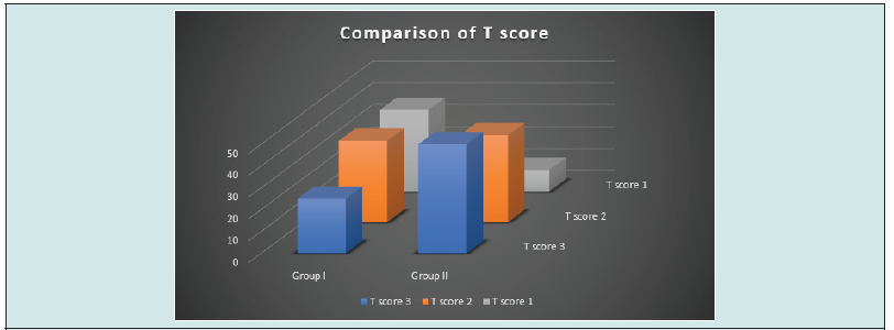
In all patients, there were 45 (56.3%) females and 35 (43.7%) males. (Figure 1) Mean age of the patients in group I was 33.3±8.35 years and in group II mean age was 34.9±7.23 years. Among 40 cases of group I, 22 (55%) patients had urban residency and in group II 19 cases had urban residency. (Table 1) In group I 31 (77.5%) cases had adequate length of RCF and in group II 32 (80%) had adequate length of RCF. As per homogeneity of RCF, 28 (70%) cases had adequate and 30 (75%) had adequate homogeneity. We found a significant difference of tapper of RCF in both groups, 13 (32.5%) cases of group I had adequate tapper while in group II 31 (77.5%) cases had adequate tapper. (Table 2) As per T score, in group II 20 (50%) cases had score 3 and 16 (40%) cases had score 2 while in group I 10 (25%) cases had score 3 and 15 (37.5%) cases had T scare 2 with p value <0.005. (Figure 2) Figure 2: T-score by using chi-square test.
Discussion
The dental community has a major concern about the prevalence of premature loss of primary molars. If you want to save your face, a pulpectomy is the best course of action. An integral part of doing a pulpectomy is cleaning and contouring the root canal. Adequate mechanical debridement and obturation quality are critical to the effectiveness of an endodontic operation. [11] Primary teeth have been used in numerous in vitro investigations comparing various rotary instrumentation devices to manual instrumentation. [12] In the current study 80 patients of both genders were presented. There were 45 (56.3%) females and 35 (43.7%) males. Patients were equally categorized in two groups. The mean age of the patients in group I was 33.3±8.35 years and in group II mean age was 34.9±7.23 years. Among 40 cases of group I, 22 (55%) patients had urban residency and in group II 19 cases had urban residency. These findings were comparable to the previous studies. [13,14] Single-rooted teeth (incisors, canines, premolars) were used in this study to evaluate the radiographic obturation quality of two root canal preparation methods (rotary vs conventional meth-od). When comparing root canal fillings completed using manual instrumentation against rotary equipment, no significant differences were seen in duration between the two groups. It seems that the proportion of properly filed cases was comparable across the two groups. In the current study, researchers found no significant difference in filling consistency between the two groups. Compared to the conventional group, the rotary group saw a significantly higher frequency of instances with sufficient density (manual). However, in a 2011 research by Robia G, it was found that the incidence of instances with acceptable density was observed to be considerably greater in the rotary group compared to the manual group. [15] This study found no statistically significant differences between the two groups (manual vs. rotary) on any of the obturation quality parameters except for taper of the root canal filling. The canal’s natural curve and taper must be preserved throughout preparation, from the canal opening to the apical foramen. [16] A larger percentage of patients in the rotary group had an acceptable taper than in the conventional group (77.5% vs. 32.5%) in this research. Using a T-score to measure the overall quality of obturation, we found a statistically significant (p 0.005) difference between the two groups. The percentage of instances with a T-score of 3 (classified as having perfect obturation) was significantly higher in the rotary group (50%) than in the manual group (25%) [17,18].
Conclusion
According to the results of this research, the rotary approach yielded higher quality obturations in terms of taper and overall quality than did the traditional method. The quality of root canal filling is an important factor in the success of an apicectomy. A wellfilled root canal prevents the recurrence of infection and promotes the healing process. The use of rotary files for root canal filling has been shown to provide better results compared to manual filling. Rotary files allow for more accurate filling of the root canal, resulting in better outcomes. Despite this, there was no difference between rotary and manual instrumentation in terms of the radiographic technical quality of the root canal obturation with respect to length and density.
References
- Chauhan A, Saini S, Dua P, Mangla R (2019) Rotary Endodontics in Pediatric Dentistry: Embracing the New Alternative. Int J Clin Pediatric Dent 12(5): 460-463.
- Ghaderi F, Jowkar Z, Tadayon A (2020) Caries Color, Extent, and Preoperative Pain as Predictors of Pulp Status in Primary Teeth. Clin Cosmet Investig Dent 12: 263-269.
- Duncan HF, Galler KM, Tomson PL, Simon S, El-Karim I, et al. (2019) European Society of Endodontology position statement: Management of deep caries and the exposed pulp. Int Endod J 52(7): 923-934.
- Burch JG, Hulen S (1972) The relationship of the apical foramen to the anatomic apex of the tooth root. Oral Surg. Oral Med. Oral Pathol. 34(2): 262-268.
- Chugal NM, Clive JM, Spångberg LSM (2003) Endodontic infection: some biologic and treatment factors associated with outcome. Oral Surg. Oral Med. Oral Pathol. Oral Radio and Endod 96(1): 81-90.
- Saunders WP, Saunders EM, Sadiq J, Cruickshank E (1997) Technical standard of root canal treatment in an adult Scottish sub-population. British Dental Journal 182(10): 382-386.
- Boltacz- Rzepkowska E, Pawlicka H (2003) Radiographic features and outcome of root canal treatment carried out in the Łodz′ region of Poland. International Endodontic Journal 36(1): 27-32.
- Sjögren U, Hägglund B, Sundqvist G, Wing K (1990) Factors affecting the long term results of endodontic treatment. Journal of Endodontics 16(10): 498-504.
- Román-Richon S, Faus-Matoses V, Alegre Domingo T, Faus-Llácer VJ (2014) Radiographic technical quality of root canal treatment performed ex vivo by dental students at Valencia University Medical and Dental School, Spain. Medicina Oral, Patología Oral Cirugía Bucal 19(1): 93-97.
- Makarem A, Ravandeh N, Ebrahimi M (2014) Radiographic assessment and chair time of rotary instruments in the pulpectomy of primary second molar teeth: a randomized controlled clinical trial. J Dent Res Dent Prospects 8(2): 84-89.
- Tabassum S, Khan FR (2016) Failure of endodontic treatment: The usual suspects. Eur J Dent 10(1): 144-147.
- Crespo S, Cortes O, Garcia C, Perez L (2008) Comparison between rotary and manual instrumentation in primary teeth. J Clin Pediatr Dent 32(4): 295-298.
- Robia G (2014) Comparative radiographic assessment of root canal obturation quality: manual verses rotary canal preparation technique. Int J Biomed Sci 10(2): 136-142.
- Govindaraju L, Jeevanandan G, Subramanian EMG (2017) Comparison of quality of obturation and instrumentation time using hand files and two rotary file systems in primary molars: A single-blinded randomized controlled trial. Eur J Dent 11(3): 376-379.
- Robia G (2014) Comparative radiographic assessment of root canal obturation quality: manual verses rotary canal preparation technique. Int J Biomed Sci 10(2): 136-142.
- Yilmaz A, Karagoz-Kucukay I (2017) In vitro comparison of gutta-per-cha-filled area percentages in root canals instrumented and obturated with different techniques. J Istanb Univ Fac Dent 51(2): 37-42.
- Casaña Ruiz, MD, Martínez LM, Miralles EG (2022) Update in the Diagnosis and Treatment of Root Canal Therapy in Temporary Dentition through Different Rotatory Systems: A Systematic Review. Diagnostics 12(11): 2775.
- Patil S, Parkarwar P (2018) Quality of root canal filling in manually and rotary-filed single-rooted teeth undergoing apicectomy in tertiary care hospital of South Punjab. Journal of Oral Biology and Craniofacial Research 8(1): 30-33.

Top Editors
-

Mark E Smith
Bio chemistry
University of Texas Medical Branch, USA -

Lawrence A Presley
Department of Criminal Justice
Liberty University, USA -

Thomas W Miller
Department of Psychiatry
University of Kentucky, USA -

Gjumrakch Aliev
Department of Medicine
Gally International Biomedical Research & Consulting LLC, USA -

Christopher Bryant
Department of Urbanisation and Agricultural
Montreal university, USA -

Robert William Frare
Oral & Maxillofacial Pathology
New York University, USA -

Rudolph Modesto Navari
Gastroenterology and Hepatology
University of Alabama, UK -

Andrew Hague
Department of Medicine
Universities of Bradford, UK -

George Gregory Buttigieg
Maltese College of Obstetrics and Gynaecology, Europe -

Chen-Hsiung Yeh
Oncology
Circulogene Theranostics, England -
.png)
Emilio Bucio-Carrillo
Radiation Chemistry
National University of Mexico, USA -
.jpg)
Casey J Grenier
Analytical Chemistry
Wentworth Institute of Technology, USA -
Hany Atalah
Minimally Invasive Surgery
Mercer University school of Medicine, USA -

Abu-Hussein Muhamad
Pediatric Dentistry
University of Athens , Greece

The annual scholar awards from Lupine Publishers honor a selected number Read More...


