Lupine Publishers Group
Lupine Publishers
Menu
ISSN: 2637-4749
Research Article(ISSN: 2637-4749) 
Cottonseed Cake Effect on Hematological, Biochemical and Histological Changes in Liver and Testes in Rabbits Volume 5 - Issue 3
Farrah Deeba1*, Anas Sarwar Qureshi2, Liaqat Ali1, Asad Niaz Khan1 and Muhammad Usman3
- 1Department of Clinical Medicine and Surgery, Faculty of Veterinary Science, University of Agriculture, Pakistan
- 2Department of Anatomy, Faculty of Veterinary Science, University of Agriculture, Pakistan
- 3Department of Basic Sciences, KBCMA, College of Veterinary and Animal Sciences, Narowal, Sub campus UVAS-Lahore, Pakistan
Received: February 14, 2023; Published: February 22, 2023
Corresponding author: Farrah Deeba, Department of Clinical Medicine and Surgery, Faculty of Veterinary Science, University of Agriculture, Faisalabad, Pakistan
DOI: 10.32474/CDVS.2023.05.000211
Abstract
Cottonseed cake contains a toxic substance gossypol. Presently cottonseed cake is widely used as a rich protein supplement to feed livestock extensively by ignoring toxicity risk. The current study is planned to observe toxic effect of feeding cottonseed cake on hematological, biochemical and histological changes in liver and testes in male rabbits. Administration of different levels of cotton seed cake (0%,10%,15%) for eight weeks induced non- significant changes in red blood cell, count (RBCs), white blood cell count (WBCs), hemoglobin (Hb), packed cell volume (PCV and platelet count (Plt). Likewise, serum transaminases alanine aminotransferase(ALT) and , aspartate aminotransferase(AST) have shown non-significant changes but serum testosterone was significantly decreased., Histopathology of liver and testes revealed non- significant changes in liver architecture but significant damage in testes tissue Therefore it is concluded that cottonseed cake feeding in rabbits produced no toxic effect on hematology and liver function but severe toxic effects on reproductive system by decreasing serum testosterone level and inducing damage in testes.
Keywords:Testes; platelet; testosterone; hemoglobin
Highlight
a) Complete blood picture of rabbits fed cottonseed cake.
b) Serum liver transaminases and liver histology of rabbits fed cottonseed cake.
c) Serum testosterone level and testes histology of rabbits fed cottonseed cake.
Introduction
Cottonseed cake, known as a rich source of protein, is used worldwide in the form of a supplement in the livestock ration to increase productivity. Cottonseed cake contains a high percentage of proteins (40-46%) and fiber. It is relatively a cheap protein source and comparable to other protein sources used as supplement in livestock including soyabean and groundnut cake. However, use of cottonseed cake as supplement in both ruminants and simple stomach animals is limited due to presence of toxic substance. Gossypol in cottonseed and cotton seed derived products [1]. Cotton (Gossyp ium spp.) pigment glands produce various phenols that are toxic specifically gossypol [2,2-bi(8–formyl-1,6,7–tri–hydroxy–5–isopropyl– 3-methylnaphthalene)]. Gossypol is present in all parts of the cotton plant, but its high levels are found in the cotton plant seeds. Livestock is prone by gossypol toxicity due to consumption of cotton plant by-products procured from processing of cotton plant fiber and cotton seeds, including cotton seed cake and meal [2].
Continuous and prolonged feeding of cottonseed by-products causes negative impact on growth performance and fertility in livestock. Common clinical signs of gossypol toxicity in both premature and mature ruminants are same and include dyspnea, weight loss, anorexia, emaciation and death after prolonged feeding [3]. Scientific literature reported that cotton seed by-products have also caused hematological alterations in ruminants and prolonged feeding might be considered dangerous . Studies further reported that free gossypol in cotton seed by-products binds to iron (Fe) causing loss of Fe and nonavailability for normal process of biosynthesis of hemoglobin and might be interacting directly on erythrocyte membrane resulting in anemia and increase in erythrocyte fragility. Therefore, consumption of cotton seed by-products has been associated with changes in hematology in ruminants [4]. Prolonged supplementation with cotton seed by-products has also caused immunosuppression and an increase in rate of various infections in animals. Moreover, changes in hematology might result from prolonged exposure to gossypol containing cotton seed by-products. Therefore, a study was carried out to estimate changes after feeding cottonseed cake at various percentage on hematology, biochemical parameters and histology of liver and testes in male rabbits.
Materials and Methods
Study design
Twelve apparently healthy male rabbits of local breed weighing approximately 1500-1800 g were considered for the current study. All rabbits were purchased from the local market. The rabbits were distributed in 3 groups randomly containing four biological replicates in one group. The rabbits were provided with seasonal fodder and fresh tap water ad libitum. Live body weight of rabbits was measured weekly. The animals were kept in the animal house of the Department of Clinical Medicine and Surgery, University of Agriculture- Faisalabad for acclimatization in the maintained environmental conditions The study was carried out by following the guidelines of the Directorate of Graduate Studies and Institutional Animal Ethical Committee. All the study groups were provided with diet Group- I (Control ): (Diet with 0% Cotton seed cake ): Group- 2(Diet with 5% Cotton seed cake);Group- 3 (Diet with 15% Cotton seed cake) for eight weeks.
Hematological and Serum Analysis
Before slaughtering the rabbits blood samples (5ml) were taken from jugular vein and shifted to vacutainers with anticoagulant (EDTA) for complete blood count (CBC) and without anticoagulant for serum collection. Serum was harvested by centrifugation at 1500 revolution per minute (rpm) for 10 minutes and stored at -20° C till further analysis. The hematological profile including red blood cell count (RBCs), white blood cell count (WBCs) hemoglobin level (Hb), packed cell volume (PCV), and platelets (Plts) were determined by following the procedure mentioned in literature [5]. Cobas C111 fully automated chemistry analyzer utilized for estimation of liver alanine aminotransferase (ALT) and aspartate aminotransferase (AST), and Backman coulter analyzer was used for estimation of serum testosterone by Chemiluminescent immunoassay method.
Histological Analysis
Rabbits were humanely slaughtered under gaseous anesthesia. All rabbits were opened for liver and testes tissue collection. Liver tissue was transferred to fixative (neutral buffered formalin) and testes were transferred to Bouin’s solution immediately after rinsing with normal saline [6]. Paraffin embedding technique was used on the tissue samples to prepare 5-micron thick sections that were subjected to Hematoxylin and Eosin (H&E) staining procedure. Slides were examined under microscope at 100X to observe degenerative changes in liver and testes.
Statistical Analysis
The data obtained were analyzed by Analysis of variance (ANOVA) technique using SPSS version 22 statistical computer software at 5% probability.
Results
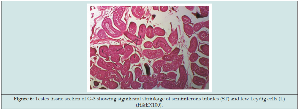
Table 1 shows changes in body weight of rabbits in all study groups from 1st week to 8th week. No toxicity signs were observed in rabbits during the study period. Table 2 shows changes in the blood parameters including red blood cells (RBCs), white blood cells (WBCs), hemoglobin concentration (Hb), packed cell volume (PCV), and platelets counts (Plts) in all study groups. Table 3 shows biochemical analysis of serum including alanine aminotransferase (ALT). aspartate aminotransferase (AST), and testosterone level in all study groups. The histomicrograph of liver tissue in G-1 appeared to be normal arrangement of hepatocytes around the central hepatic vein (Figure 1). In G- 2 liver tissue revealed non-significant damage to liver cells with minimum infiltration of inflammatory cells around the central hepatic vein (Figure 2). In G-3 liver tissue revealed non-significant liver cell disruption and derangement of hepatocytes around the central hepatic vein (Figure 3). Histomicrograph of testes tissue of G-1 revealed normal cellular structure of seminiferous tubules with Leydig cells and interstitial tissues (Figure 4). Histomicrograph of testes of G-2 presented shrinkage in the testicular seminiferous tubules (Figure 5). Histomicrograph of testes in G-3 presented shrinkage of the testicular seminiferous tubules and the cellular distance was significantly increased with few Leydig cells (Figure 6).
Different superscripts in the same column mean significant difference at a significant level (P<0.05).
Different superscripts in the same column mean significant difference at a significant level (P<0.05).
Different superscripts in the same row mean significant difference at a significant level (P<0.05).
Figure 1: Liver tissue section of G-1 showing normal hepatic cord (hc) and normal portal triad (PT) (H&EX100).
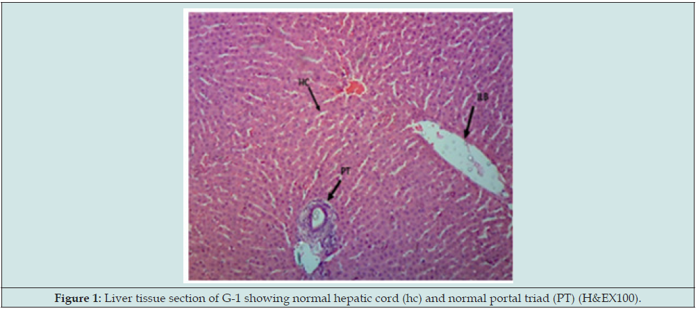
Figure 2: Liver tissue section of G-2 showing non- significant damage in hepatocytes(h) and Inflammatory cell infiltration (If) (H&EX100).
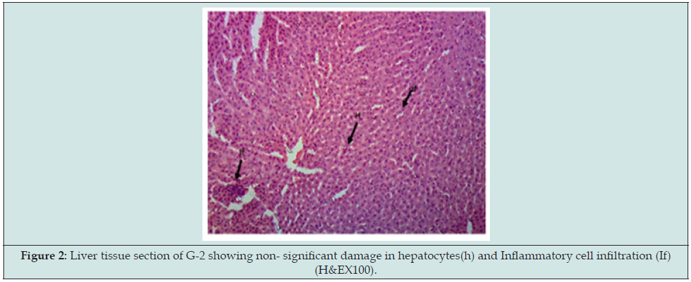
Figure 3: Liver tissue section of G-3 showing non-significant damage in hepatocytes(h) and Inflammatory cell infiltration (If) (H&EX100).
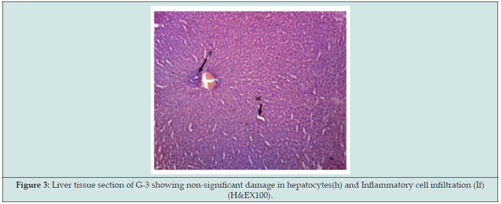
Figure 4: Testes tissue section of G-1 showing normal pattern of seminiferous tubules with normal Leydig cells(L) (H&EX100).
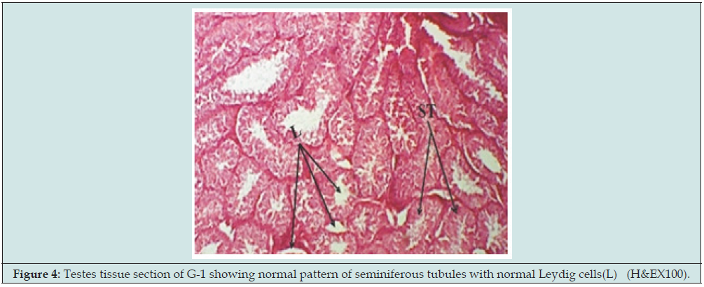
Figure 5: Testes tissue section of G-2 showing significant shrinkage of seminiferous tubules (ST) and few Leydig cells (L) (H&EX100).
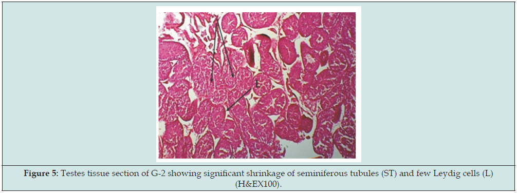
Figure 6: Testes tissue section of G-3 showing significant shrinkage of seminiferous tubules (ST) and few Leydig cells (L) (H&EX100).
Discussion
In concurrence with the increasing trend of supplementing cottonseed cake in livestock diet, various studies were accomplished explaining the effects of toxic substance gossypol present in cotton by-products affecting different body systems. In the current study the body weight of rabbits in all study groups increased significantly the possible reason might be the diet provided with all essential nutrients along with an acceptable level of protein thus rabbits were able to utilize them effectively for growth. Similar findings were reported by Amao et al. [7] they reported that cotton seed level in diet has non- significant effect on growth and performance of male rabbits as measured by weekly gain in body weight, final gain in body weight ,weekly consumption of feed with feed efficiency. The current findings are in contrast to Taha et al. [8] they observed that high concentration of gossypol in diet of rabbits caused decrease in body weight as compared to control group. Hematological findings in our study showed non- significant change in red blood cell in contrast to study by Al Tayyar et al. [9] where significant decrease in red blood cell count was observed after treatment with gossypol for 10 days in male carp. The decline was attributed to the alteration in the structure, function and integrity of erythrocyte membrane by direct binding of gossypol.
Total white blood cell count indicates functionality of immune mechanism of the body and is closely associated to humoral and cellular immunity. The findings of our study showed non- significant alteration in white blood cells in contrast to the study by Al-Tayyar et al. who reported a decrease in white blood cells count and attributed it to destruction of blood forming organs in case of gossypol toxicity. Moreover, Akingbemi et al. [10] stated that gossypol is associated with malnutrition leading to anemia, leucopenia and thrombocytopenia due to a depression in bone marrow activity in rats El-Mokadem et al. [11] further explained the possible mechanism of toxicity by gossypol as inhibition of glucose-6-phosphate dehydrogenase resulting in reduced production of NADPH, needed for the activity of an essential cellular antioxidant named glutathione peroxidase. Decreased activity of the enzyme as a part of cellular antioxidant system might initiate an excessive deposition of cellular oxidants causing decrease in packed cell volume and hemoglobin levels. Similarly, Lindsey et al. [12] observed alterations in fragility of erythrocytes and Hb and attributed these physiological changes to feeding cotton seed meal in dairy cows. The absence of literature in non-ruminants indicates that etiology of gossypol intoxication in ruminants might be different from ruminants.
Current study shows non- significant changes in ALT and AST in all study groups. Our findings are in alignment with Camara et al. who observed no consistent changes in ALT, AST, γ-glutamyl transferase and concluded that feeding cotton seed cake in sheep did not lead to liver or kidney injury. Current findings are in contrast to Akingbemi et al. stated that gossypol significantly increased serum ALP and ALT activities in rats. Al Sharaky et al. [13] also observed that liver transaminases (ALT and AST) and alkaline phosphatase (ALP) increased at a significant level in rats treated with gossypol . The increase in serum transaminases indicate cellular damage predominantly in hepatocytes including muscle cells. In the present study blood serum testosterone level in G-3 were significantly low as compared to G-1. Our findings are consistent with El-Mokadem et al. They found reproductive toxicity due to presence of gossypol in sheep and concluded that blood serum concentration of testosterone reduced due to presence of gossypol in diet. Moreover Taha et al. Evaluated the effect of two dose levels (4 and 20mg/ kg body weight) of gossypol on alternate days on quality of semen and hormonal profile of male rabbits for four months. Rabbits treated with gossypol have reduced blood plasma levels of testosterone accompanied by decline in sperm concentration, functional sperms fraction and initial concentration of fructose in semen.
Gu et al. [14] explained that in cattle decreased steroidogenesis in corpus luteum cells in gossypol treated group might be associated with enzymes function adenylate cyclase and 3α-hydroxysteroid dehydrogenase. Donaldson et al. [15] reported an in vitro study and concluded that gossypol inhibited testosterone production by dispersion of Leydig cells in rodents. They observed significant increase in the activity of 17β-HSD and 17-ketostroid decreased in gossypol treated animals that might be a possible explanation for reduction in serum testosterone levels. In the present study non -significant changes were observed in the liver architecture in all study groups. Our findings are in contrast to Wang and Lei [16] where feeding gossypol acetic acid to rats manifested marked histopathological alterations in liver cells including vacuolization in mitochondria, dilation of endoplasmic reticulum , widening of peri-nuclear space with proliferation of collagen fibers extensively in the Disse’s space .They further stated that gossypol binds to microsome protein in an irreversible manner both in presence or absence of electron donor NADPH and thus concluded that gossypol acetic acid has potential to cause damage to liver cells. Similar findings were reported by Akingbemi et al. They found vacuolar degeneration of hepatocytes in gossypol treated rats. Dearas et al. [17] reported gossypol as a potent toxic agent which can induce severe liver damage including biliary hyperplasia, peripheral deposition of iron pigments in the liver of young ruminants.
They further stated that gossypol deposited in high concentration in liver cells. The liver generally appeared pale with distinct pinpoint foci of coagulative necrosis. In the present study significant shrinkage of seminiferous tubules with increase in cellular distance and very few Leydig cells were observed in G-3. Similarly, Yan Chang [18] reported that germ cell damage in the seminiferous tubules epithelium was observed after in vivo administration of gossypol. In contrast to our findings Hassan et al. [19] reported that gossypol treated bulls showed non- significant effect on the motility of sperms, testes morphometry and histological architecture of the testes. The toxic effect of feeding gossypol on morphology of spermatozoa was reversible. Feeding gossypol for fifty-six days showed significant abnormalities in sperms but the abnormalities were reversible. Hoffer [20] Studied the effects of various levels of gossypol for eleven weeks on rat testes. Under the light microscope, both extensively damaged and entirely normal seminiferous tubules placed close to one another in the same tissue. Damage in seminiferous tubules manifested as intraepithelial vacuolization of different sizes, extensive exfoliation and atrophy of structures Moreover, electron microscope observations manifested intraepithelial vacuole formation. Severely damaged Sertoli cells exhibited various large vacuoles and decline in cytoplasm ground substance, both types of endoplasmic reticulum and also golgi apparatus. These changes were manifested as early as 2 weeks after gossypol administration and increased significantly with an increase in dose of gossypol and feeding time.
Conclusion
The present study suggested that supplementing various levels of whole cotton seed cake for a short time showed no adverse effects on hematological parameters and liver function but induced histopathological changes in testes in rabbits.
Acknowledgments
The authors are grateful to the faculty of Veterinary Science for helping to achieve this work.
Conflict of interest
The authors declare no conflict of interest regarding the publication of this paper.
References
- Phyu HW, Po SP, Hein ST, Khaing KT, Win MM, et al. (2017) A study on the effects of cottonseed cake on histopathological changes of liver in sheep. Int J Vet Sci Anim Husb 2(4): 10-13.
- Gadelha ICN, Fonseca NBS, Oloris SCS, Melo MM, Soto-Blanco B (2014) Gossypol toxicity from cottonseed products. Sci World J 2014: 231635.
- Randel RD, Chase CC, Wyse ST (1992) Effects of gossypol and cottonseed products on reproduction of mammals. J Anim Sci 70(5): 1628-1638.
- Camara ACL, Menezes do vale A, Mattoso CRS, Melo M, Soto-Blanco B (2016) Effects of gossypol from cottonseed cake on the blood profile in sheep. Trop Anim Health Prod 48(5):1037-1042.
- Mohammed IH, Kakey ES (2020) Effect of Prosopis farcta extracts on some complications (hematology and lipid profiles) associated with alloxan induced diabetic rats. Iraqi J Vet Sci 34(1): 45-50.
- Qassim AH, Al- Sammak MA, Ayoob AA (2022) Histological changes in kidney and pancreas induced by energy drinks in adult male rats. Iraqi J Vet Sci 36(1): 111-116.
- Amao OA, Adejumo DO, Togun VA, Oladunjoye IO (2012) Growth response and nutrient digestibility of pre-pubertal rabbit bucks fed cotton seed cake-based diets supplemented with vitamin E. Afr J Biotech 11(76): 14102-14109.
- Taha TA, Shaaban WF, El Mahdy AR, El Nouty FD, Salem MH (2006) Reproductive toxicological effects of gossypol on male rabbits: semen characteristics and hormonal levels. Ani Sci 82(2): 259-269.
- Al Tayyar OBA, Al-Beiati MA, AL Ubade SA. (2009) Gossypol effect in some blood parameters enzymes and pathology of male common carp Cyprinus carpiol. Iraqi J Vet Sci 33(1): 173-179.
- Akingbemi BT, Aire TA (1994) Haematological and serum biochemical changes in the rat due to protein malnutrition and gossypol ethanol interactions. J Comp Pathol 111(4): 413-426.
- El Mokadem MY, Taha TA, Samak MA, Yasen AM (2012) Alleviation of reproductive toxicity of gossypol using selenium supplementation in rams. J Anim Sci 90(9): 3274-3285.
- Lindsey TO, Hawkins GE, Guthrie LD (1980) Physiological responses of lactating cows to gossypol from cottonseed meal rations. J Dairy Sci 63(4): 562-573.
- El-Sharaky AS, Newairy AA, Elguindy NM, Elwafa AA (2010) Spermatotoxicity, biochemical changes and histological alteration induced by gossypol in testicular and hepatic tissues of male rats. Food Chem Toxicol 48(12): 3354-3361.
- Gu Y, Lin YC, Rikihisa Y (1990) Inhibitory effects of gossypol steroidogenic pathways in cultured bovine luteal cells. Biochem Biophys Res Commun 169(2): 455-461.
- Donaldson A, Sufi SB, Jeffcoate SI (1985) Inhibition by gossypol of testosterone production by mouse Leydig cells in vitro. Contraception 31(2): 165-71.
- Wang Y, Lei HP (1987) Hepatotoxicity of gossypol in rats. J Ethanopharmacol 20(1): 53-64.
- Deoras DP, Young-Curtis P, Dalvi RR, Tippett FE (1997) Effect of gossypol on hepatic and serum gamma-glutamyl transferase activity in rats. Vet Res Commun 21(5): 317-323.
- Yan Chang C, Mruk DD (2002) Cell junction dynamics in the testis: Sertoli-germ cell interactions and male contraceptive development. Physiol Rev 82(4): 825-874.
- Hassan ME, Smith GW, Ott RS, Faulkner DB, Firkins LD, et al. (2004) Reversibility of the reproductive toxicity of gossypol in peri pubertal bulls. Theriogenology 61(6): 1171-1179.
- Hoffer AP (1983) Effects of gossypol on the seminiferous epithelium in the rat: a light and electron microscope study. Biol Reprod 28(4): 1007-1020.

Top Editors
-

Mark E Smith
Bio chemistry
University of Texas Medical Branch, USA -

Lawrence A Presley
Department of Criminal Justice
Liberty University, USA -

Thomas W Miller
Department of Psychiatry
University of Kentucky, USA -

Gjumrakch Aliev
Department of Medicine
Gally International Biomedical Research & Consulting LLC, USA -

Christopher Bryant
Department of Urbanisation and Agricultural
Montreal university, USA -

Robert William Frare
Oral & Maxillofacial Pathology
New York University, USA -

Rudolph Modesto Navari
Gastroenterology and Hepatology
University of Alabama, UK -

Andrew Hague
Department of Medicine
Universities of Bradford, UK -

George Gregory Buttigieg
Maltese College of Obstetrics and Gynaecology, Europe -

Chen-Hsiung Yeh
Oncology
Circulogene Theranostics, England -
.png)
Emilio Bucio-Carrillo
Radiation Chemistry
National University of Mexico, USA -
.jpg)
Casey J Grenier
Analytical Chemistry
Wentworth Institute of Technology, USA -
Hany Atalah
Minimally Invasive Surgery
Mercer University school of Medicine, USA -

Abu-Hussein Muhamad
Pediatric Dentistry
University of Athens , Greece

The annual scholar awards from Lupine Publishers honor a selected number Read More...


