Lupine Publishers Group
Lupine Publishers
Menu
Case Report(ISSN: 2770-5447) 
Coronary Cameral Fistulae a Scarce Entity Volume 1 - Issue 1
Ranjan Modi1*, Prabhu Halkati2 and Suresh Patted3
- 1Associate consultant, Fortis Escorts Heart Institute, India
- 2,3Kle Hospital, India
Received: January 18, 2018; Published: January 31, 2018
Corresponding author: Ranjan Modi, Associate consultant, Fortis Escorts Heart Institute, India
DOI: 10.32474/ACR.2018.01.000101
Abstract
Coronary artery fistulae are communication between coronary arteries and other structures like cardiac chamber (coronary cameral fistula) or a vein (coronary arteriovenous fistula). Coronary fistulae account for 0.2 to 0.4% of the congenital cardiac abnormalities. We present a 20 day old baby with coronary cameral fistula of the right coronary ostium which was draining into the right ventricle then right atrium and into the pulmonary artery.
Keywords: Coronary fistulae; Congenital abnormality; Cameral fistulae
Case Report
Coronary artery fistulae are communication between coronary arteries and other structures like cardiac chamber (coronary cameral fistula) or a vein (coronary arteriovenous fistula) [1]. Coronary fistulae account for 0.2 to 0.4% of the congenital cardiac abnormalities. Spontaneous closure occurs in 23% of small fistulae, primarily those arising from left coronary system in which conservative management may be appropriate [2]. Though surgery still remains the main stay of treatment, interventional closure seems to be an appropriate choice. We present a 20 day old male baby born to non consangious marriage. The neonate was full term delivery in the hospital. Prenatal history was uneventful with no history of any drug intake during pregnancy and regular antenatal care visits with complete immunizations. The baby presented with history of feeding difficulty since birth, no history of recurrent chest infections, no suck rest suck cycle and no cyanosis. The patient had tachycardia with heart rate of 140/min and tachypnoea with respiratory rate of 36/min on examination, blood pressure of 90/60 mmHg.
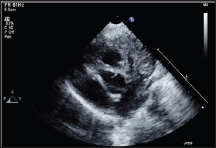
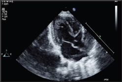
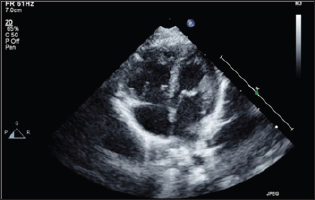
Examination of the cardiovascular system revealed normal first and second heart sounds with continuous murmur in the the precordium. Transthoracic Echocardiography revealed situs solitus of d loop ventricles, short axis and parasternal long axis revealed well visualized left coronary ostia but an aneurysmal dilatation of the right coronary ostia (Figures 1 & 2), dilated right atrium (RA) right ventricle (RV) and dilated pulmonary artery (Figures 3 & 4). The patient underwent coronary angiography through the right transfemoral route which revealed aneurysmal dilatation of the right coronary ostium which was draining into the right ventricle then right atrium and into the pulmonary artery (Figure 5). The patient was advised percutaneous coil closure of the fistula but the parents refused for any intervention. The patient was discharged against medical advice.
Figure 1: 2D Echocardiography short axis showing the presence of left coronary ostia and dilated right coronary ostia.

Figure 2:2D Echocardiography parasternal long aixs showing dilated Right coronary cusp

Figure 3: 2D Echocardiography Apical Four chamber view revealed dilated RA/RV.

Figure 4: 2D Echocardiography revealed dilated pulmonary artery.

Figure 5: Coronary angiography revealed ectasia of the right coronary artery ostiae draining into the right ventricle to right atrium and pulmonary artery

Discussion
Coronary fistulae involve communications between a coronary artery with a cardiac chamber (coronary cameral fistula) or a vein (coronary arteriovenous fistula). The coronary fistulae are rare and 60% arise from the right coronary artery. The majority (>90%) of cameral fistulae drain in the right-sided chambers of the heart and the rest into the left side of the heart or to both. The majority of these patients present in adulthood and are usually asymptomatic, most often being detected accidentally. They appear as a persistence of the sinusoidal connection between the lumen of the primitive tubular heart which supplies the myocardial blood flow in the early embryonic period. The path physiology of coronary fistula is myocardial stealing or decrease in myocardial blood flow distal to the site of connection [3]. The coronary vessel tries to compensate for the reduction in coronary blood flow by progressive enlargement of the ostia and the feeding artery. The management of cameral fistulae is uncertain due to the rarity of the condition. Surgery is the main stay of treatment, in a review done [4], 57% of patients were treated surgically with a 0.5% operative mortality rate, with complications including myocardial ischaemia (3%) and recurrence (4%).
Interventional catheterization is yet another therapeutic option available for treatment. The technique uses coils or other devices and can be performed on an outpatient basis [5]. advocated intra-cardiac closure of the fistula as external plication technique had higher recurrence rates. Herein is reported a rare condition of coronary cameral fistula of right coronary artery draining into the right ventricle .The rarity is exponentiated due to the presence of this condition in a 20 day old baby. Various treatments options, interventional and surgical, were available for the management of the baby but sadly could not be undertaken due to unavailability of consent from the parents. Even though adequate management was not possible in this baby, the case report was with notifying in view of its rarity.
References
- Yamanaka O, Hobbs RE (1990) Coronary artery anomalies in 126,595 patients undergoing coronary arteriography. Cathet Cardiovasc Diagn 21(1): 28-40.
- Mahoney LT, Sckieken RM, Rauer RM (1982) Spontaneous closure of a coronary artery fistula in childhood. Pediatr Cardiol 2: 311-312
- Fyfe DA, Edwards D, Driscoll DJ (1986) Myocardial ischemia in patients with pulmonary atresia and intact ventricular septum. J Am Coll Cardiol 8(2): 402-406.
- McNamara JJ, Gross RE (1969) Congenital coronary artery fistula. Surgery 9: 59.
- Cheung DL, Au WK, Cheung HH, Chiu CS, Lee WT (2001) Coronary artery fistulas: long-term results of surgical correction. Ann Thorac Surg 9: 190-195.

Top Editors
-

Mark E Smith
Bio chemistry
University of Texas Medical Branch, USA -

Lawrence A Presley
Department of Criminal Justice
Liberty University, USA -

Thomas W Miller
Department of Psychiatry
University of Kentucky, USA -

Gjumrakch Aliev
Department of Medicine
Gally International Biomedical Research & Consulting LLC, USA -

Christopher Bryant
Department of Urbanisation and Agricultural
Montreal university, USA -

Robert William Frare
Oral & Maxillofacial Pathology
New York University, USA -

Rudolph Modesto Navari
Gastroenterology and Hepatology
University of Alabama, UK -

Andrew Hague
Department of Medicine
Universities of Bradford, UK -

George Gregory Buttigieg
Maltese College of Obstetrics and Gynaecology, Europe -

Chen-Hsiung Yeh
Oncology
Circulogene Theranostics, England -
.png)
Emilio Bucio-Carrillo
Radiation Chemistry
National University of Mexico, USA -
.jpg)
Casey J Grenier
Analytical Chemistry
Wentworth Institute of Technology, USA -
Hany Atalah
Minimally Invasive Surgery
Mercer University school of Medicine, USA -

Abu-Hussein Muhamad
Pediatric Dentistry
University of Athens , Greece

The annual scholar awards from Lupine Publishers honor a selected number Read More...


